Blefaroplasma® with PLASMAGE® may be defined as a non invasive eyelid treatment that improves the abnormal function, reconstructs deformities or
enhances appearance and may be either reconstructive or cosmetic (aesthetic)
Dermatochalasis, including symptomatic redundant skin weighing down on the upper eyelashes (i.e. pseudoptosis) and surgically induced dermatochalasis after prosis repair.
Acquired blepharoptosis, may result from streching, dehiscence, or disinsertion of the levator aponeurosis. Aponeurotic blepharoptosis is commonly known as involutional ptosis in patients in which
the anatomic changes are age-related.
Brow ptosis, drooping of the eyebrows to such an extent that excess tissue is pushed into the upper eyelid.
The plasma generated by the ionization of the gaz creates a sublimation of superficial tissues thus creating a lifting effect.
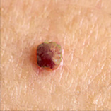
Cutaneous fibroma is a relief or a skin growth of normal skin color springs, is typically connected to the skin by a stalk. The skin papilloma may occur in any area of the body, although the preferred locations are the areas where there are skin folds such as the eyelids and around the eyes, the sides of the neck, armpits, groin and upper of the chest.These small skin tumors are benign and are usually asymptomatic unless they are not traumatized voluntarily or involuntarily by clothes or other.
There are no known causes, however, it seems that the irritation due to rubbing in skin folds stimulates growth. The pendulous fibroids are very common in middle age. They develop in both men and women. They may be skin-colored or darker and of variable size between 1 and 5 mm.
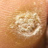
Verruca Vulgaris - A flesh-colored, firm papule or nodule due to infection of epidermal cells with human papillomaviruses. Also known as warts.
On close inspection, normal skin lines over the surface of the lesion are typically disrupted. The dome-shaped lesions can also be studded with black puncta. The growth is characterized by hypertrophy of dermal papillae and thickening of the keratin layers of the epidermis. The surface is hyperkeratotic with many small filamentous projections. Verrucae commonly occur on hands and fingers, and can occur in groups or in a linear pattern.
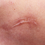
Hypertrophic Scars are characterized by excessive deposition of collagen in the dermis and subcutaneous tissues secondary to traumatic or surgical injuries.
Contrary to the asymptomatic fine-line scar that results from normal wound repair, the exuberant scarring of hypertrophic scars results typically in distressing disfigurement, hypertrophic scars are erythematous.
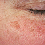
A lentigo is a small, sharply circumscribed, pigmented macule surrounded by normal-appearing skin. Histologic findings may include
hyperplasia of the epidermis and increased pigmentation of the basal layer. A variable number of melanocytes are present; these melanocytes may be increased in number, but they do not form nests.
Lentigines may evolve slowly over years, or they may be eruptive and appear rather suddenly. Pigmentation may be homogeneous or variegated, with a color ranging from brown to black.
Multiple clinical and etiologic varieties exist. The distinction of a lentigo from other melanocytic lesions (eg, melanocytic nevi, melanoma) and its role as a marker for ultraviolet damage and
systemic syndromes is of major significance.
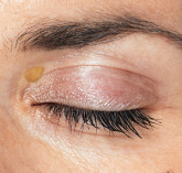
Xanthelasma palpebrarum is the most common form of xanthoma.
The lesions appear as yellowish, flat, soft, with different form and dimension, are located mostly at the medial angle of the eyelid. It is usually bilateral and is characterized by the development
of yellowish plaques related to the presence of cholesterol.
Lesions are initially situated in the medial canthus and gradually spread to all of the periorbital region in advanced forms.
Histological examination reveals esterified cholesterol deposits situated in the cytoplasm of histiocytes in the middle and superficial layers of the dermis and epidermis.


